Keratoconus
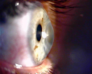
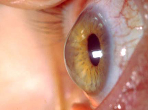
Keratoconus is a degenerative and progressive back of the eye disease, which is manifested by the development konus forms of corneal stromal thinning and its protrusion. In nearly all cases the disease is bilateral, and as a rule the clinical picture is more pronounced in one eye.
The first sensations associated with the occurrence of diseases usually occur during puberty or during the twenties. Diagnostic procedures that are necessary for the diagnosis and monitoring the progression of keratoconus, in addition to a standard ophthalmologic examination, is the examination of corneal topography with Oculyzer, which very accurately register the slightest changes in corneal curvature and thinning stromal tissue.
The above-mentioned structural changes that result in corneal distortions manifest objects in the visual field, duplicated images with the advent of 'ghost images' and the general reduction in visual acuity of patients eye.
In the initial stages of keratoconus, satisfactory visual acuity can be achieved by adjusting the glasses, but particularly successful GP cone contact lenses.
However, if the disease progression is evident, as a solution imposed by the surgical approach, in the form of intact intra-corneal rings, corneal cross-linking and penetrating keratoplasty as the final solution.
At the clinic MILMEDIC we perform method of cross-linking / CXL / drops with the use of riboflavin and UV lamps, with the aim of strengthening the corneal stromal matrix.
Extensive experience of the surgical team led by Prof. Dr. M.Vukosavljević is a guarantee of good postoperative results.